Retina Unit
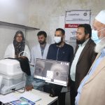
Retina is the innermost coat of the eyeball responsible for image perception. Medical retina service at Mazharul Haque BNSB Eye Hospital deals with the patients having retinal disorders that can be managed medically without any surgical intervention. The medical retina clinic is equipped with modern facilities like Fundus Camera, fluorescein angiography, Optical coherence tomography and all types of laser services. This clinic is also running a project named “Setup Retina Unit” which has been set-up in collaboration with ANDHERI HILFE, eV, Bonn, Germany. In this project, patient with retinal disorder and diabetic retinopathy are screened, graded & treated accordingly through invasive and non invasive procedures. This clinic also provides counselling to the patients & training for the doctors. Besides, through tele-medicine this service is connected to the other centres at home & abroad.
Surgical retina service at this hospital deals with the patients having retinal disorders that require surgical intervention. The retinal surgeons perform around 100 surgeries per year of a variety of vitreoretinal conditions like retinal detachment, Intraocular foreign body removal, vitreous haemorrhage, proliferative vitreoretinopathy, advanced diabetic eye disease etc.
Management of Mazharul Haque BNSB Eye Hospital were grateful to ANDHERI HILFE for their generoug contribution to setup Retina Unit at the hospital. We are also thankful to the donners, partners and supporters of ANDHERI HILFE, Bonn, Germany.
Patients Screening Statistics
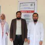
This specialized Retina Unit Established in November 2022 at offer variety of services to treat Retinal disorder and Diabetic Retinopathy at affordable cost.
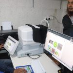
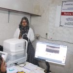
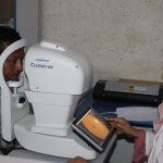
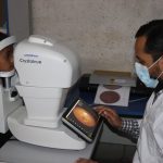
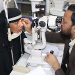
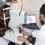
OCT Examination Area in Retina Unit of Mazharul Haque BNSB Eye Hospital. Thank you ANDHERI HILFE for supporting high Tech Medical Equipment’s for the poor and ultra poor community of Bangladesh
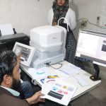
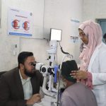
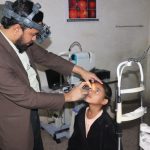
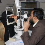
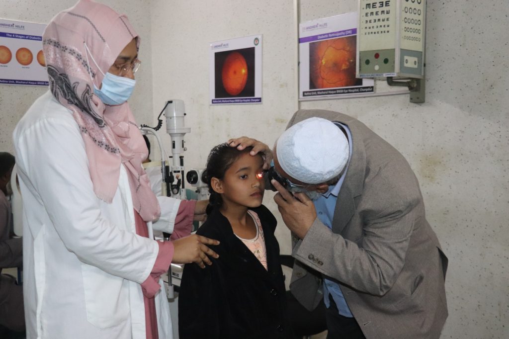
Fundus Examination Area in Retina Unit of Mazharul Haque BNSB Eye Hospital. Thank you ANDHERI HILFE for supporting high Tech Medical Equipment’s for the poor and ultra poor community of Bangladesh
Specialized Retina Unit
Mazharul Haque BNSB Eye Hospital established the first Vitreo-Retina (VR) Clinic in Chandpur and its associated areas in 2022. It is one of the major Advance secondary care eye hospitals receiving referral cases from all over surrounding districts. The clinic has state-of-the-art equipment for investigation and treatment of various Vitreo retinal diseases.
Diagnosis & Treatment
The clinics are well equipped with trained doctors and modern technology to diagnose and treat a multitude of vitreo retinal conditions. Treatment includes both medical and surgical interventions.
State-of-the-art equipment for investigation and diagnosis of various Vitreo retinal diseases include:
- Fundus Fluorescein Angiography (FFA)
- Optical Coherence Tomography (OCT)
- Ultrasonography
- Electroretinogram (ERG)
The Vitreo-Retina clinics offer medical management for various retinal conditions like Diabetic Retinopathy, retinal vein occlusion (CRVO & BRVO), Arterial occlusion (CRAO), Central serous retinopathy (CSR), Age-related Macular Degeneration (AMD) etc., The clinics are equipped with retinal lasers (Green laser – Double frequency YAG) with slit-lamp and indirect ophthalmoscopy delivery for treatment of these retinal conditions. In addition, various intravitreal injections of Anti-VEGF drugs and steroids are used to treat macular edema as well as choroidal neovascular membranes (CNVM). Specialized lasers like trans pupillary thermotherapy (TTT) and photodynamic therapy (PDT) are also available.
The Vitreo-Retina clinics are involved in screening for Retinopathy of Prematurity (ROP) at base clinics as well as NICUs in other hospitals. Various pediatric retinal conditions like FEVR, PFV, tumors etc are also treated regularly.
The Vitreo-Retina clinics have operating theatres equipped with state-of-the-art vitrectomy machines, lasers and microscopes with posterior visualisation systems. Complex vitreo-retinal surgeries for repair of retinal detachment, diabetic tractional detachment, vitreous haemorrhage, macular holes and epiretinal membranes are routinely performed. Surgeries to manage intra-ocular foreign bodies and treatment of endophthalmitis are also routinely done.
Retinal Diseases
- Diabetic Retinopathy
- Macular Degeneration
- Retinal Detachment
- Retinopathy of Prematurity
- Vein Occlusions
- Intravitreal Injections
- Laser Treatment (Photocoagulation)
- Vitrectomy
Laser Treatment
During laser treatment, a laser is used to close leaky blood vessels in the eye. It may be used to treat:
- Diabetic retinopathy
- Retinopathy of prematurity
- Macular degeneration
- Retinal vein occlusions
During treatment:
- Your eyes will be temporarily numbed with local anesthetic eye drops
- Eye drops are used to widen your pupils
- A contact lens is used to hold your eyelids open
- The laser beam is focused on your retina
The entire procedure normally takes 20-40 minutes, so you should not have to stay in the hospital overnight. You may need to come back for another round of laser treatment. Because of the anesthetic, the treatment should not be painful.
Beyond the Retina Unit
Diabetic Retinopathy is an eye condition caused by diabetes. In diabetes, high sugar levels can damage the retina by slowly weakening blood vessels in the eye. At first, this causes no symptoms. However, without treatment, it can cause blindness.
Diabetic Retinopathy can take years to cause blindness. In the early stages, it is treatable. If you are a diabetic, visit your eye doctor every year. Early detection is very important and can save your eyesight!
Intravitreal injections are drugs that are injected into the eye to reduce swelling and capillary growth. Usually, the drugs belong to the “Anti-Vascular endothelial growth factor” group of medicines, so the treatment may be referred to as “Anti-VEGF” treatment. However, in some cases, steroid drugs may be injected instead.
Conditions Treated with Intravitreal Injections:
- Macular degeneration (specifically Wet Age-related Macular Degeneration)
- Diabetic Retinopathy
- Retinal Vein Occlusions
Many retinal disorders involve abnormal or damaged blood vessels. These vessels may leak blood into the surrounding area, resulting in gradual vision loss. With laser treatment, the high energy light beam seals the capillaries shut. Closing the blood vessels may not restore your vision. But it can prevent the blood from leaking out of the capillaries. This stops the condition from getting any worse.
During laser treatment, a laser is used to close leaky blood vessels in the eye. It may be used to treat:
- Diabetic retinopathy
- Retinopathy of prematurity
- Macular degeneration
- Retinal vein occlusions
Vitrectomy is an eye surgery used to treat retinal diseases. It involves the clear, jelly-like fluid (vitreous) that fills the back of the eye. The vitreous gives shape to the eyeball just like air gives shape to a balloon. But in some diseases, it becomes cloudy or starts pulling on the retina. In these cases, the vitreous may need to be replaced. Vitrectomy is the surgery used to remove and replace this fluid.
Conditions treated with vitrectomy:
- Vitreous hemorrhage
- Severe eye trauma
- Diabetic Retinopathy
- Retinal Detachment
- Retinopathy of Prematurity
Retina is the light sensitive layer of tissue that lines the inside of the eye and sends visual messages through the optic nerve to the brain. When the retina detaches, it is lifted or pulled from its normal position. If not treated promptly, retinal detachment can cause permanent vision loss.
Key Points to Remember:
- If you have any sudden change in vision, see an eye doctor immediately!
- Low Vision Clinics an help you adjust to a life with low vision
Macular Degeneration is a condition that causes central vision loss. Central vision is what your eyes focus on when you look straight ahead. Macular Degeneration only affects central vision because it is caused by damage to the central part of the retina (called the macula). It does not affect side vision.
Retinopathy of Prematurity (ROP) occurs due to abnormal growth of blood vessels in an infant’s eye. During development, blood vessels grow from the central part of the retina outwards. This process is completed few weeks before the normal time of delivery. However in premature babies, it is incomplete. If blood vessels grow normally, ROP does not occur. On the contrary, if the vessels grow and branch abnormally the baby develops ROP.
The light-sensitive tissue in your eye is called the retina. It gives us the ability to see because it is made up of nerve cells that detect light. Normally, these nerve cells get nutrients from your arteries and dump waste into your veins. But if a vein is blocked, it cannot carry blood away from the retina. Instead, fluid leaks out of the vein. This is called a vein occlusion.
Causes & Risk Factors:
Retinal veins are very narrow. If a large clot tries to pass through, it can block the vein, causing vein occlusion.
The risk of vein occlusion is higher for those with:
- Diabetes
- High blood pressure
- High cholesterol levels
- Health problems that affect blood flow
For Accessible, Affordable and Quality Eye Care Service please contact with us. You can also support our project and activities for the most vulnerable community of Bangladesh.

